
Sep 12 • Nakyoung Lee
[Lecture Review] [MRI Online] Knee MRI Part 2.
The continuation of the CME series, MRI-Based Anatomy of the Knee: Cruciate & Collateral Ligaments with Patellofemoral Stabilizers, provides an advanced dissection of knee MRI anatomy. This lecture dives into the cruciate system (ACL & PCL), the layered complexity of the medial collateral ligament (MCL), and the medial patellofemoral ligament (MPFL), translating fine structural anatomy into clinically relevant insights.
Lecture Overview – This lecture is structured around the following key learning objectives:
1. Identify MPFL/MPML, MCL layers (SMCL, TCL, POL), and ACL/PCL on MRI.
1. Identify MPFL/MPML, MCL layers (SMCL, TCL, POL), and ACL/PCL on MRI.
2. Assess patellofemoral alignment and cruciate integrity across planes.
3. Distinguish true ligament tears from common mimics.
From Layers to Ligaments: MRI as a Structural Roadmap
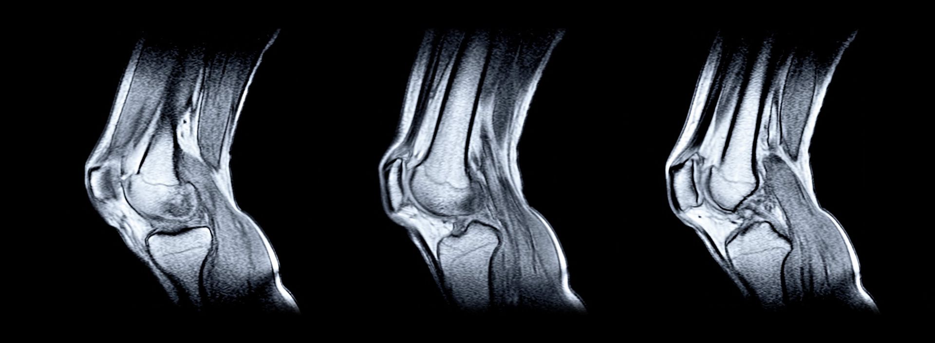
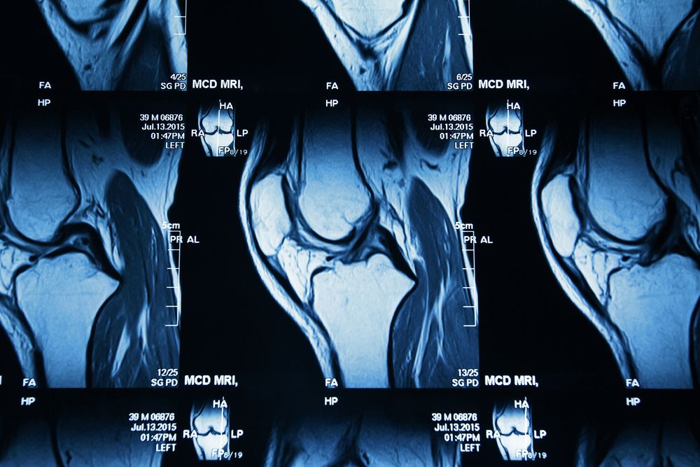
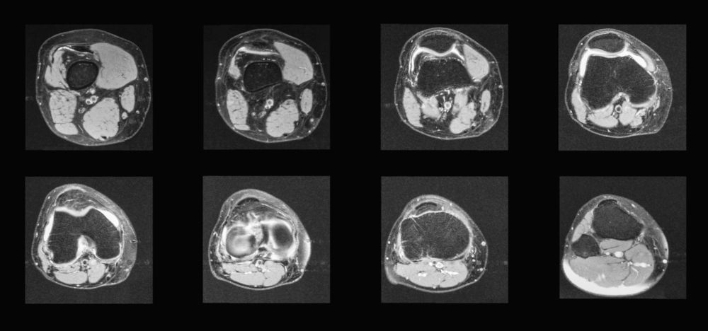
This lecture emphasizes not only ligament visualization but also the interplay of supporting tissues and how their injury translates to pathology. Multiple MRI planes—sagittal, axial, and coronal—are used to show the dynamic relationship of bundles, footprints, and sheaths.
Key highlights include:
Anterior Cruciate Ligament (ACL) :
The ACL is shown as a two-bundled structure (anteromedial and posterolateral), likened to a “celery stalk.”
The ACL is shown as a two-bundled structure (anteromedial and posterolateral), likened to a “celery stalk.”
The over-the-top femoral attachment landmark and fanning at the tibial insertion are clearly demonstrated.
Pitfalls such as pseudomasses from synovial sheaths and false impressions from fiber twisting are explained, with coronal and axial views rescuing diagnostic ambiguity
Posterior Cruciate Ligament (PCL) :
Posterior Cruciate Ligament (PCL) :
Described as thicker, arc-shaped, and prone to interstitial rather than complete tears.
Bundle anatomy (anterolateral vs posteromedial) is linked to flexion–extension mechanics.
Meniscofemoral ligaments (Humphrey & Wrisberg) are highlighted as key stabilizers of the lateral meniscus, with cautionary notes on pseudolesions and pitfalls like pseudo–bucket handle tears
Medial Collateral Ligament (MCL) :
Presented through Warren & Marshall’s three-layer model (layers 1–3), clarified to avoid common terminology confusion.
Presented through Warren & Marshall’s three-layer model (layers 1–3), clarified to avoid common terminology confusion.
Layer 1: Crus fascia and medial retinaculum.
Layer 2: Superficial MCL (historically “tibial collateral ligament”).
Layer 3: Capsular layer, including meniscotibial and meniscofemoral ligaments, blending into the posterior oblique ligament (POL) and oblique popliteal ligament (OPL).
Axial and coronal MRI views illustrate how these layers fuse, separate, and contribute to joint stability.
Medial Patellofemoral Ligament (MPFL) :
Carefully distinguished from surrounding retinacular fascia, correcting the common misconception of being synonymous with the medial retinaculum.
Medial Patellofemoral Ligament (MPFL) :
Carefully distinguished from surrounding retinacular fascia, correcting the common misconception of being synonymous with the medial retinaculum.
Its role in preventing lateral patellar dislocation is emphasized, with insertion traced between the adductor tubercle and medial femoral epicondyle.
Integration with the superficial MCL and POL at a “global train station” demonstrates the complexity of patellofemoral stabilization.
Why This Lecture Matters
For clinicians managing knee instability—whether cruciate tears, MCL sprains, or patellar dislocations—this lecture provides both the anatomic depth and MRI-based confidence needed for accurate interpretation and intervention planning. It moves beyond textbook structures, showing real imaging nuances, pitfalls, and clinical correlations.
Ready to Elevate Your MRI Knee Interpretation?
Dive into this lecture to strengthen your command of cruciate and collateral anatomy, sharpen your recognition of subtle stabilizers like the MPFL, and refine your approach to diagnosing complex injuries.
An indispensable session for orthopedic surgeons, sports medicine physicians, musculoskeletal radiologists, and integrative clinicians.
What to read next on Jaseng Medical Academy
Get in touch
-
536, Gangnam-daero, Gangnam-gu, Seoul, Korea
-
jaseng.education@jaseng.co.kr
-
+82 2 2222 2792
-
Contact Us
Family Sites
Our Newsletter
Receive Jaseng Medical Academy's updates and events
Thank you!

Copyright © 2019 - 2026 Jaseng Medical Academy. All rights reserved.