Lecture Overview – This lecture is structured around the following key learning objectives:
1. Identify major ligaments and tendons of the knee on MRI and understand their anatomical roles.
2. Assess patellofemoral and tibiofemoral alignment and its implications for joint function.
3. Recognize key neurovascular structures and their relevance in knee injuries.
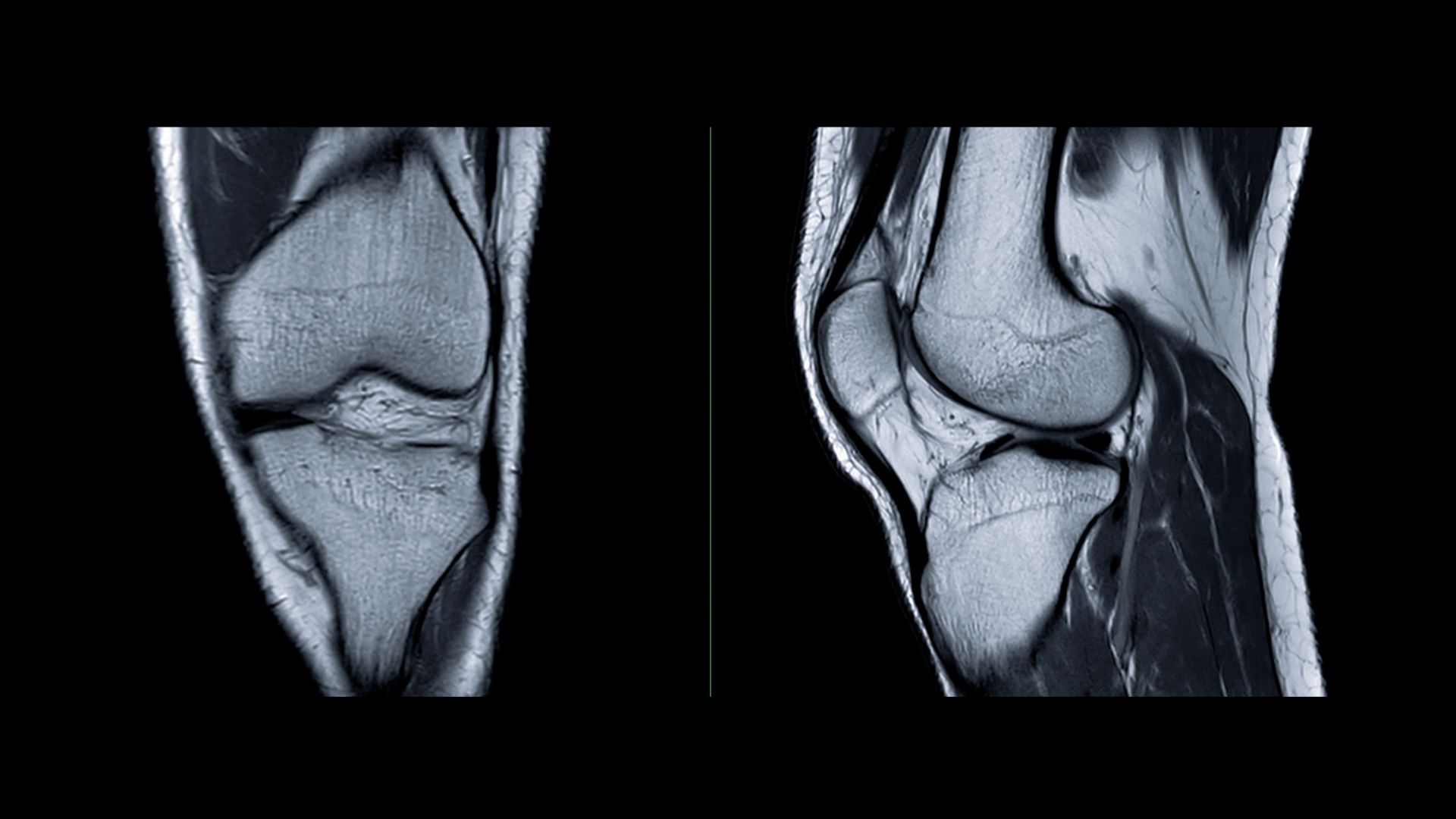
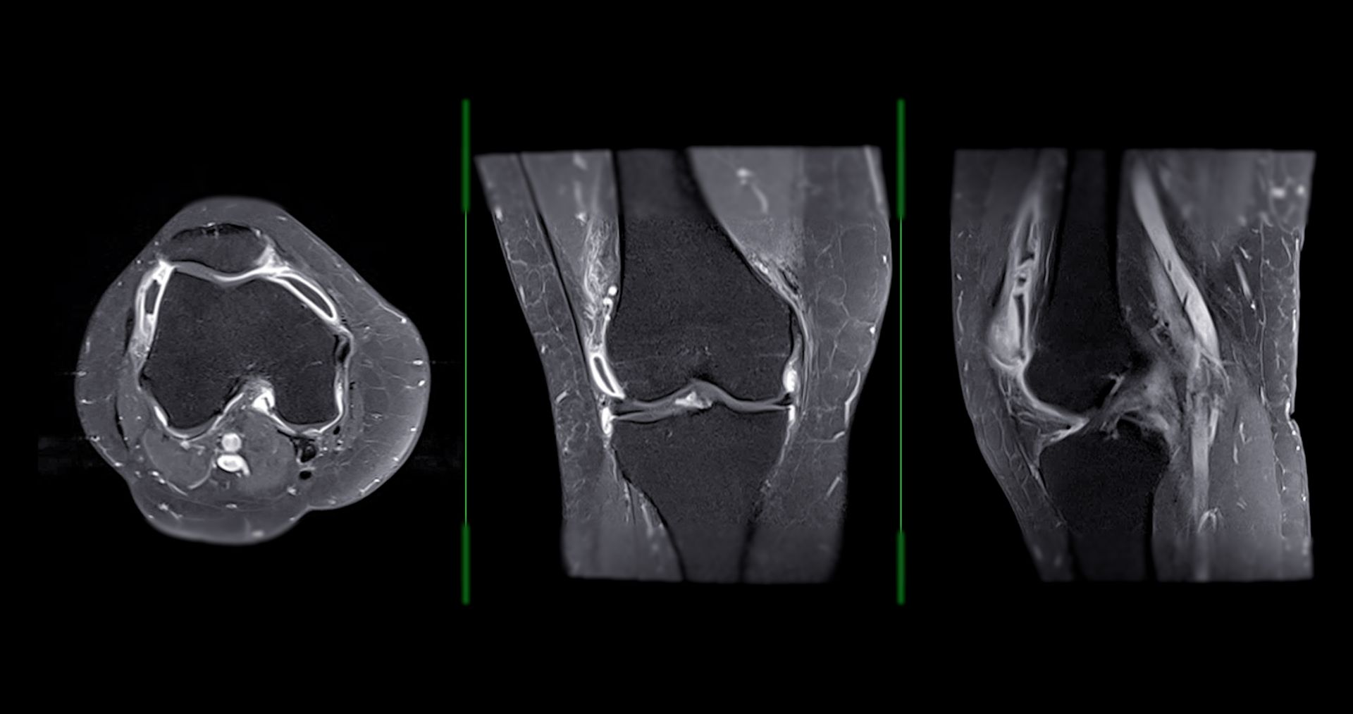
This lecture stands out by walking the viewer through the knee in multiple MRI planes—axial, sagittal, and coronal—highlighting how anatomy and clinical pathology intersect.
Key highlights include:
Cruciate Ligaments (ACL & PCL):
Their orientation, insertion sites, and stabilizing functions are visualized with clarity. Important accessory structures such as the ligaments of Humphrey and Wrisberg are also covered, providing deeper insights into cruciate function and surgical visibility.
Collateral Ligaments (MCL & LCL):
The lecture explains the three-layered anatomy of the medial collateral complex, including the often-overlooked posterior oblique ligament. Lateral support structures such as the fibular collateral ligament and biceps femoris tendon are shown with practical notes on varus and valgus stability.
Tendons & Soft Tissue Anatomy:
The pes anserinus group, semimembranosus expansions, quadriceps, and patellar tendon anatomy are covered in depth, with MRI-based guidance for identifying their insertions and recognizing signs of overuse or injury.
The popliteal fossa is explored in detail, emphasizing the relative depth of the artery, vein, and tibial nerve—critical for evaluating trauma and detecting pathologies like thrombosis or adventitial cysts.
The course also offers a nuanced look at the medial patellofemoral ligament (MPFL) and its role in patellar stabilization, correcting common misconceptions about its relationship with the medial retinaculum.
For clinicians navigating complex knee pathologies—from ligament injuries to neurovascular entrapments—this lecture provides both anatomical clarity and interpretive confidence.
Whether you're refining your MRI reading skills or planning targeted interventions, the content bridges radiological detail with clinical utility.
Watch the full lecture to deepen your understanding of knee structure and function through the lens of high-resolution MRI.
It's an essential tool for orthopedic specialists, musculoskeletal radiologists, physical medicine practitioners, and integrative medicine clinicians.